![]()
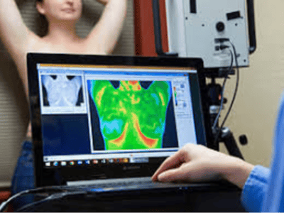
Thermography is simply a procedure utilizing an electronic camera to obtain an image of the infrared radiation (heat) coming from the surface of the skin. It is performed as an aid to the diagnosis of abnormal temperature patterns, which may or may not indicate the presence of a disease process or pathology.
Medical research has shown thermography to be helpful in the identifying:
- Peripheral Nervous System Disorders
- Metabolic Disorders
- Repetitive Strain Injuries
- Headaches, Neck and Back Problems
- TMJ Conditions
- Dental & Sinus Infections
- Pain Syndromes such as fibromyalgia, myositis
- ArthritisVascular Disorders (ex. Raynoud’s disease)
- Soft Tissue Injuries
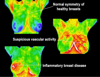
There are patterns detectable by thermograms that suggest any condition for which there is an alteration of nervous control of blood flow or circulation such as soft tissue injuries, diabetes, peripheral vascular disease, osteoarthritis, dental and sinus infections, breast implant rupture, strokes, thyroid inflammation or underfunction, gall-bladder and liver inflammation, lymph congestion, and melanoma.
Clinical correlation is always required
 The appointment takes place in a relaxed, peaceful & private room. The room air may feel cool as we adjust to room temperature before scanning. You will be lightly robed during the cooling process (about ten minutes). During the examination you will disrobe in areas being scanned for both imaging purposes and to allow for the surface temperature of the body to acclimate with the room.
Obtaining a thermographic image is like having your picture taken. There is no direct contact between you and the camera. There is no radiation, no injection, nothing to drink. The procedure is totally non- invasive and the camera does not emit radiation of any kind.
Your information is uploaded through a secure server and assigned to a medical doctor for final interpretation (similar to the process for x-rays with a radiologist). Once completed, the report is forwarded to you and any other health care practitioner requested. If you have any questions or concerns once receiving your results, we are always available to consult with you.
A color printed report and images are generally available within 3-7 business days. Your images will be read by Dr. Gregory Melvin who is trained and certified to interpret thermograms by the American College of Clinical Thermography. Dr. Melvin will analyze the results and make recommendations as to how you can go about bringing your body back into balance and prevent further dysfunction.
It is recommended to establish a baseline with the initial scan and a follow-up scan within 4 to 6 months then annually. Scans can be performed more if desired.
While participation in a DITI early detection program can increase your chance of detecting and monitoring breast disease, as with all other tests, it is still not 100% guarantee of detection.
Call 440-725-3511 to schedule.